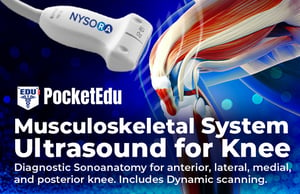
MSK for Diagnosis and Injection Therapy
Learn Ultrasound MSK of the knee in a heartbeat. Features static and dynamic examination videos. This is a highly visual, inspiring course that feels like an e-workshop. Learn VISUALLY with step-by-step videos. This course features both US anatomy and interventional regenerative therapy by the world’s leader in US-MSK for the knee. Join thousands of MSK practitioners enriching their practice with this course’s no-hassle tips on how to optimize imaging, and practical decision-making processes by world-renowned Dr. Stanley Lam.
Author: NYSORA, Dr. Stanley LamE-book content

Subscribe now and gain access to the revolutionary NOTES and Search Tools, and NEVER again loose links, articles, videos and educational images. Once you experience PocketEdu, you will never go to old books and books again.
If you want to share or recommend this product to a friend, use the below link
More products
Ban C.H. Tsui, Santhanam Suresh
Pediatric Atlas of Ultrasound and Nerve Stimulation-Guided Regional Anesthesia


Enhance your learning experience

Learn anytime, anywhere
Get 24/7 access to your learning materials on any device. Learn where, whenever, and however you like.

Stay organized
You’ll never need to go back and forth between texts and illustrations again. Mark, label favorites and take notes.

Your study materials in one place
Organize your notes, videos, links, images and attachments into your own study notebook.

Integrated search options
Use built-in PubMed and Google search engines to access the latest information on the topics of interest and save them into your notes.

Custom made illustrations
Comprehend and memorize the material faster with our proprietary illustrations and animations that will inspire you to study.

Track your progress
Keep track of where you are along your learning journey and easily pick up where you left off.