Ultrasound-Guided Cervical Plexus Block

Anesthesia and analgesia for superficial neck surgery.

Chapter Sections:


Indications: Anesthesia and analgesia for carotid endarterectomy, superficial neck, and analgesia for clavicle fractures

Carotid endarterectomy.


Goal: Local anesthetic spread around the superficial branches of the cervical plexus

Transducer: Linear

Needle: 25-22 gauge, 5 cm short bevel

Local anesthetic volume: 5-8 mL

The Cervical Plexus
The cervical plexus originates from the anterior rami of C1-C4 that combine into 3 loops from which the deep and superficial branches arise (Fig-1).

Deep (muscular) branches:

  • The phrenic nerve (C3-C5): innervates the diaphragm
  • Nerves to the geniohyoid and thyrohyoid (C1): innervate muscles and soft tissues of the airway
  • Ansa cervicalis (C1-C3): supplies muscles important for swallowing and speech

Fig-1. Anatomy of the deep cervical plexus with main branches and anastomoses.


Superficial branches:

  • Emerge from the prevertebral fascia in between the longus capitis and the middle scalene muscles to run deep to the sternocleidomastoid muscle (SCM).
  • As the SCM forms a "roof" over these branches, they eventually emerge from behind the posterior border of the muscle (Fig-2).
  • This occurs approximately at the intersection with the external jugular vein (Erb's point).

The superficial branches of the cervical plexus are:

  • Greater auricular nerve
  • Lesser occipital nerve
  • Transverse cervical nerve
  • Supraclavicular nerves

Fig-2. Superficial branches of the cervical plexus at Erb's point.

Cross-sectional anatomy

  • Cranial to C4, the branches of the cervical plexus are located within the prevertebral fascia in a groove between the longus capitis and the middle scalene muscle (Fig-3).
  • At the C4- C5 level, the branches of the cervical plexus are located between the prevertebral fascia, overlying the interscalene groove, and the investing layer of the deep cervical fascia.
  • At the C6-C7 level, the branches of the cervical plexus are located superficially to the investing layer of the cervical fascia. 

Fig-3. Transverse view of the cervical plexus at the level of C4 and C6.

Ultrasound anatomy

The cervical plexus can be visualized as small oval hypoechoic (dark) nodules immediately deep or lateral to the posterior border of the sternocleidomastoid muscle (SCM) (Fig-4).
The greater auricular nerve can be commonly seen over the SCM as a small, round, hypoechoic structure.

Fig-4. Branches of the cervical plexus (yellow arrows) at the level of C4. GaN, greater auricular nerve; SCM, sternocleidomastoid muscle; LCa, longus capitis muscle; LCo, longus Colli muscle; MSM, middle scalene muscle; LsCa, longissimus capitis muscle; LS, levator scapulae muscle; SPL, splenius capitis muscle; SECM, semispinalis capitis muscle.


An intermediate cervical plexus block provides sensory anesthesia of the anterolateral neck, retro-auricular areas, and the skin overlying and immediately inferior to the clavicle.

Sensory distribution of a cervical plexus block.
  • Patient position: Supine or semi-sitting, with the head turned away from the side to be blocked. This facilitates access to the anterolateral aspect of the neck.
  • External landmarks: Posterior border of the sternocleidomastoid muscle at the midpoint between the mastoid process and clavicle.


Patient position for an intermediate cervical plexus block.

General considerations:

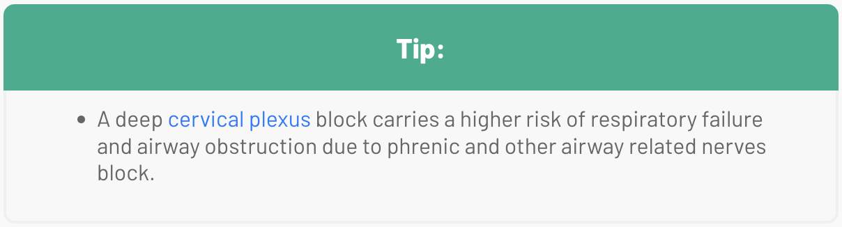
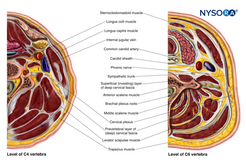
  • For a deep injection: Local anesthetic is injected at the C2-C4 level. This technique blocks the entire plexus and is generally not recommended due to the high risk of complications.
  • For an intermediate injection: Local anesthetic is injected at the C4-C5 level, between the prevertebral fascia and the investing layer of the deep cervical fascia, under the sternocleidomastoid muscle (SCM). This is the most common technique used in clinical practice.
  • For a superficial injection: Local anesthetic is injected at the level of C6, subcutaneously, superficial to the investing layer of the deep cervical fascia.

INTERMEDIATE CERVICAL PLEXUS BLOCK 
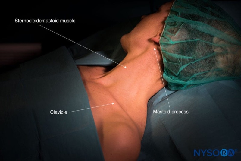
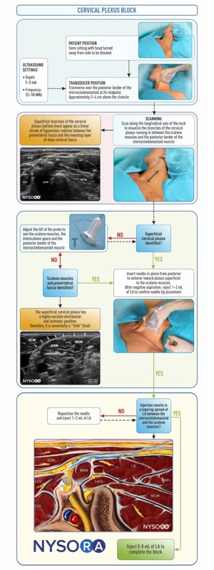
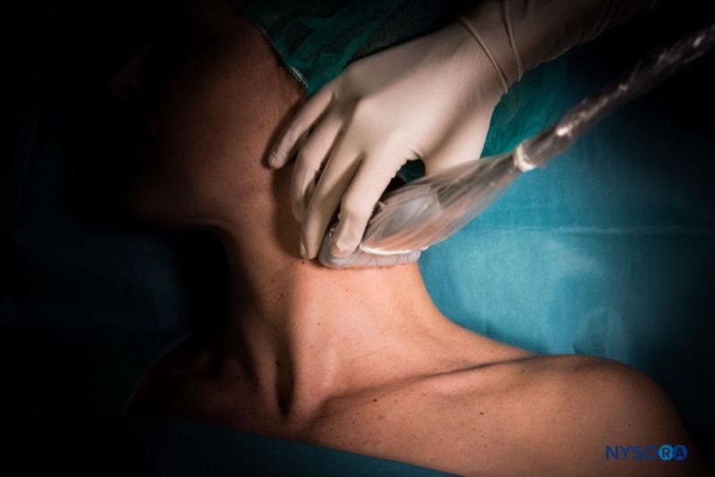
Transducer position

Transverse over the lateral aspect of the neck, overlying the SCM at the midpoint between the mastoid process and clavicle.

Scanning

  • Identify the SCM and place the posterior edge in the middle of the screen.

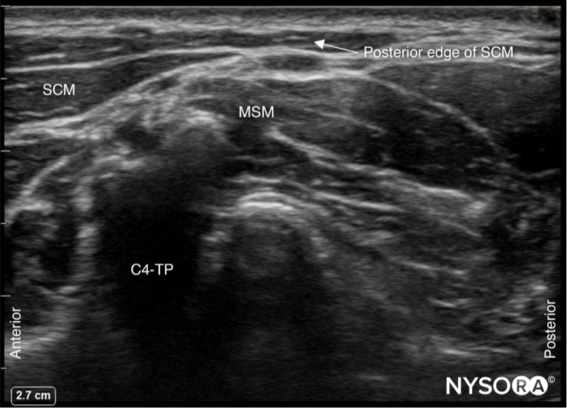
SCM, sternocleidomastoid muscle; MSM, middle scalene muscle; C4-TP, transverse process of C4.


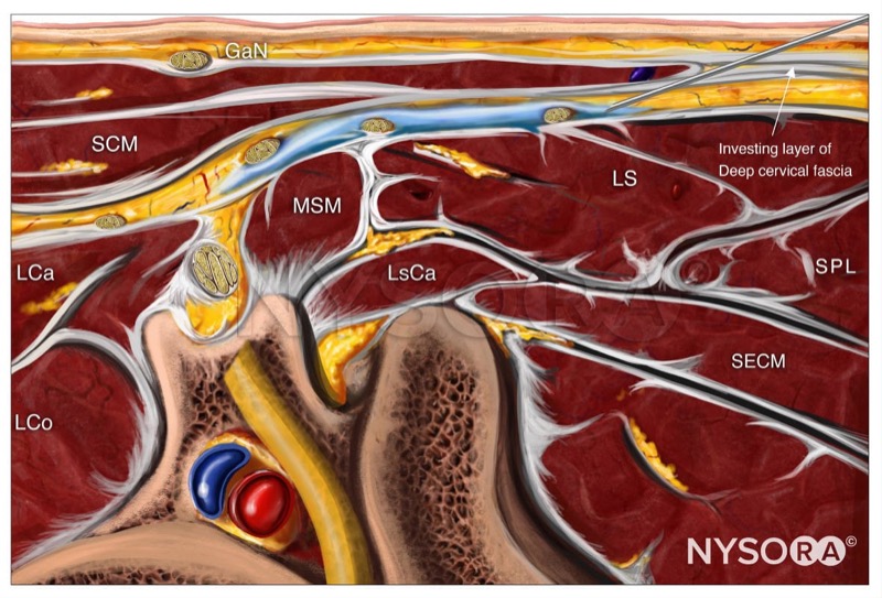
  • Slide the transducer cranial and caudally to identify the superficial branches of the cervical plexus as a small collection of hypoechoic nodules (yellow arrows) located under the SCM.

GaN, greater auricular nerve; SCM, sternocleidomastoid muscle; LCa, longus capitis muscle; LCo, longus Colli muscle; MSM, middle scalene muscle; LsCa, longissimus capitis muscle; LS, levator scapulae muscle; SPL, splenius capitis muscle; SECM, semispinalis capitis muscle.


Needle insertion

  • Insert the needle in-plane through the skin, platysma, and investing layer of the deep cervical fascia.

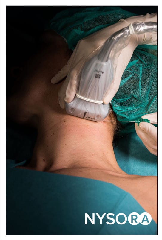
  • Position the needle tip behind the posterior border of the SCM.
  • Inject 1–2 mL of local anesthetic to confirm proper distribution between the fascial layers containing the branches of the cervical plexus.

GaN, greater auricular nerve; SCM, sternocleidomastoid muscle; LCa, longus capitis muscle; LCo, longus Colli muscle; MSM, middle scalene muscle; LsCa, longissimus capitis muscle; LS, levator scapulae muscle; SPL, splenius capitis muscle; SECM, semispinalis capitis muscle.


  • Complete the block with 5-8 mL.

Alternative transducer position

  • Place the transducer in a coronal orientation over the SCM to obtain a longitudinal view.
  • Insert the needle in-plane towards the space behind SCM, superficial to the prevertebral fascia.
  • Inject 1-2 mL of local anesthetic to confirm needle position.
  • Complete the block with a volume of 5-8 mL.

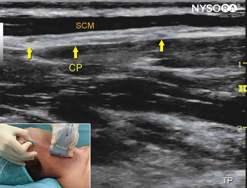
Alternative longitudinal approach for an intermediate cervical plexus block. CP, cervical plexus; SCM, sternocleidomastoid muscle.


Let's review the block: 

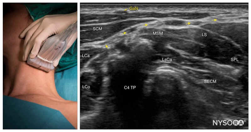
Intermediate cervical plexus; transducer position and sonoanatomy at the C4 level. GaN, greater auricular nerve; SCM, sternocleidomastoid muscle; LCa, longus capitis muscle; LCo, longus Colli muscle; MSM, middle scalene muscle; LsCa, longissimus capitis muscle; LS, levator scapulae muscle; SPL, splenius capitis muscle; SECM, semispinalis capitis muscle.


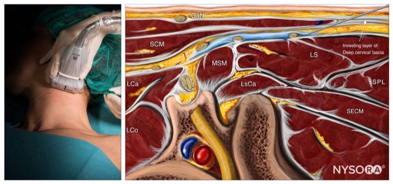
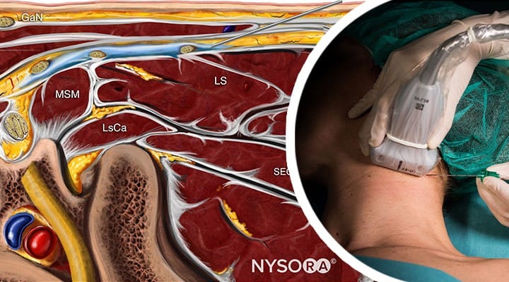
Intermediate cervical plexus block; Reverse Ultrasound Anatomy. GaN, greater auricular nerve; SCM, sternocleidomastoid muscle; LCa, longus capitis muscle; LCo, longus Colli muscle; MSM, middle scalene muscle; LsCa, longissimus capitis muscle; LS, levator scapulae muscle; SPL, splenius capitis muscle; SECM, semispinalis capitis muscle.
  • The intermediate cervical plexus block provides sensory anesthesia of the neck, without motor block.
  • Long-acting local anesthetics are typically used for carotid surgery, while short-acting agents are useful for superficial neck procedures such as node biopsy.


  • The “deep” cervical plexus block is basically a cervical paravertebral block, an advanced technique with a high risk of complications.
  • The superficial cervical plexus block technique is simpler, safer, and, for most indications adequate.
  • In-plane or out-of-plane? The superficial location of the cervical plexus branches makes both approaches possible.
  • Do I need to visualize plexus? Not necessarily or always possible.
  • Carotid surgery also requires block of the glossopharyngeal nerve branches. This block can be accomplished intraoperatively by injecting the local anesthetic into the carotid artery sheath.

GaN, greater auricular nerve; SCM, sternocleidomastoid muscle; LCa, longus capitis muscle; LCo, longus Colli muscle; MSM, middle scalene muscle; LsCa, longissimus capitis muscle; LS, levator scapulae muscle; SPL, splenius capitis muscle; SECM, semispinalis capitis muscle; LA, local anesthetic.